Top Medical Photography Techniques for Researchers
- rami7704
- Mar 17
- 4 min read
Medical photography is a vital tool in the field of research, providing visual documentation that can enhance understanding, communication, and education. Whether you are capturing images for clinical studies, educational materials, or patient documentation, mastering the right techniques can significantly improve the quality of your work. In this post, we will explore the top medical photography techniques that researchers should consider to elevate their photographic skills and produce impactful images.
Understanding the Basics of Medical Photography
Before diving into advanced techniques, it’s essential to grasp the fundamentals of medical photography. This includes understanding the equipment, lighting, and composition that contribute to high-quality images.
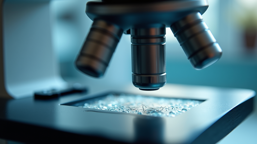
Equipment Essentials
Camera: A digital single-lens reflex (DSLR) or mirrorless camera is often preferred for its versatility and image quality. Look for models with macro capabilities for close-up shots.
Lenses: Macro lenses are crucial for capturing fine details, especially in dermatology or histology. A standard lens can work for broader shots.
Tripod: Stability is key in medical photography. A tripod helps eliminate camera shake, especially in low-light conditions.
Lighting: Natural light is ideal, but when that’s not available, consider using diffused artificial lighting to minimize harsh shadows and reflections.
Composition Techniques
Rule of Thirds: Position key elements along the lines or intersections of a grid to create a balanced composition.
Focus on Details: In medical photography, details matter. Use shallow depth of field to isolate the subject and blur the background.
Consistent Angles: Maintain consistent angles and distances when photographing similar subjects to ensure uniformity in your documentation.
Advanced Techniques for Medical Photography
Once you have a grasp of the basics, you can explore more advanced techniques that can enhance your medical photography.
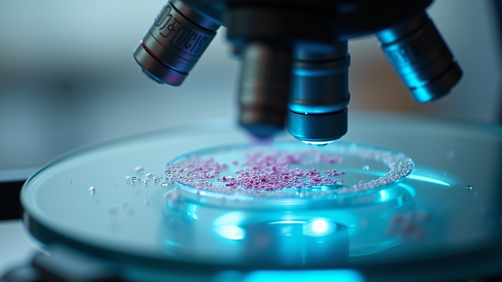
Macro Photography
Macro photography allows researchers to capture intricate details that are often invisible to the naked eye. This technique is particularly useful in fields like dermatology, pathology, and microbiology.
Use a Macro Lens: A dedicated macro lens will enable you to focus on subjects at a very close range, capturing fine details such as skin textures or cellular structures.
Lighting: Proper lighting is crucial in macro photography. Use ring lights or diffused flash to illuminate your subject evenly without creating harsh shadows.
High Dynamic Range Imaging (HDR)
HDR imaging is a technique that combines multiple exposures to create a single image with a greater dynamic range of luminosity. This is particularly useful in medical photography where both bright and dark areas need to be visible.
Capture Multiple Exposures: Take several shots at different exposure levels. Use software to merge these images into one HDR photograph.
Post-Processing: Use editing software to fine-tune the HDR image, ensuring that details in both the highlights and shadows are preserved.
Image Annotation
Annotating images can provide context and enhance understanding. This is particularly useful in educational settings or when presenting findings.
Use Software Tools: Programs like Adobe Photoshop or specialized medical imaging software allow you to add labels, arrows, and other annotations directly onto your images.
Keep it Clear: Ensure that annotations are legible and do not clutter the image. Use contrasting colors for text and arrows to make them stand out.
Ethical Considerations in Medical Photography
When engaging in medical photography, ethical considerations are paramount. Researchers must ensure that they respect patient privacy and adhere to legal guidelines.
Informed Consent
Always obtain informed consent from patients before photographing them or their medical conditions. This includes explaining how the images will be used and ensuring that patients understand their rights.
Anonymity
When publishing or sharing images, ensure that patient identities are protected. This may involve blurring faces or removing identifiable features from images.
Compliance with Regulations
Familiarize yourself with local regulations regarding medical photography. This includes understanding HIPAA guidelines in the United States or GDPR in Europe, which govern the use of personal health information.
Practical Applications of Medical Photography
Medical photography serves various purposes in research and clinical practice. Here are some practical applications:
Clinical Documentation
Photographs can serve as a permanent record of a patient’s condition, aiding in diagnosis and treatment planning. For example, documenting the progression of a wound can help assess healing over time.
Educational Materials
Images can enhance educational materials for both medical professionals and patients. High-quality photographs can illustrate complex concepts, making them easier to understand.
Research Publications
In research, photographs can provide visual evidence to support findings. Including well-captured images in publications can enhance the credibility of the research.
Conclusion
Mastering medical photography techniques is essential for researchers aiming to enhance their documentation and communication efforts. By understanding the basics, exploring advanced techniques, and adhering to ethical guidelines, you can produce high-quality images that significantly contribute to your field. Remember, the goal is not just to take pictures but to tell a story through your images. As you continue to refine your skills, consider how you can apply these techniques to your research and clinical practice, ultimately improving patient care and advancing medical knowledge.

Comments